Complex Distal Leg Ulcer
Clinical presentation, surgical treatment and documented healing.
- 01 / Problem
- Clinical presentation
- 02 / Procedure
- Surgical approach
- 03 / Outcome
- Healing & follow-up
Clinical photographs below include wounds and surgical treatment.
01 / CLINICAL PRESENTATION
Problem
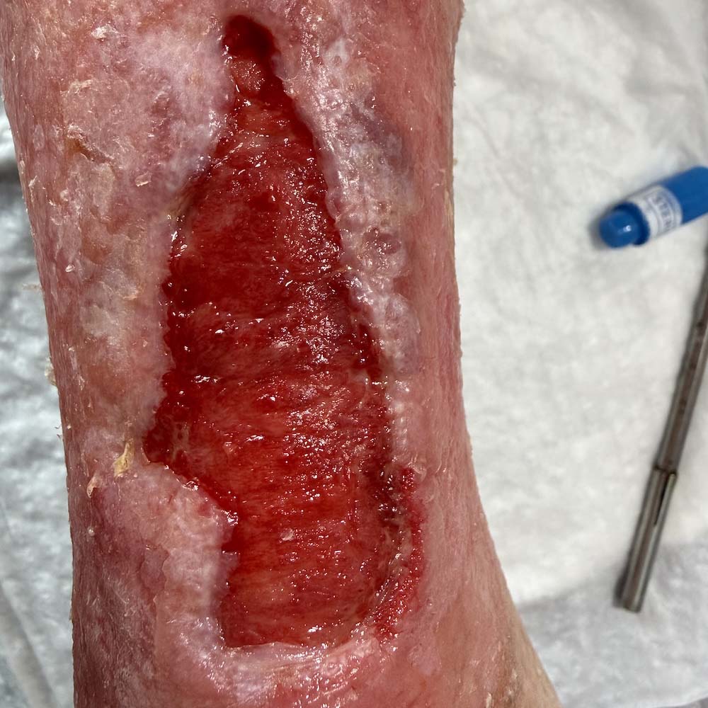
An 83-year-old woman with type 2 diabetes presented with a chronic ulcer located at the distal lower leg, extending toward the ankle—a region where wound healing is particularly challenging because of limited soft-tissue coverage, impaired vascularity, and constant mechanical stress. Despite local wound care, the ulcer failed to heal and required advanced surgical management.
02 / SURGICAL APPROACH
Procedure
Following meticulous surgical debridement, all devitalized tissue was removed to establish a healthy, well-vascularized wound bed. A split-thickness skin graft was then applied to promote definitive wound coverage and accelerate healing. Standard postoperative wound care and close clinical follow-up supported successful graft incorporation and progressive tissue regeneration.

03 / HEALING & FOLLOW-UP
Outcome
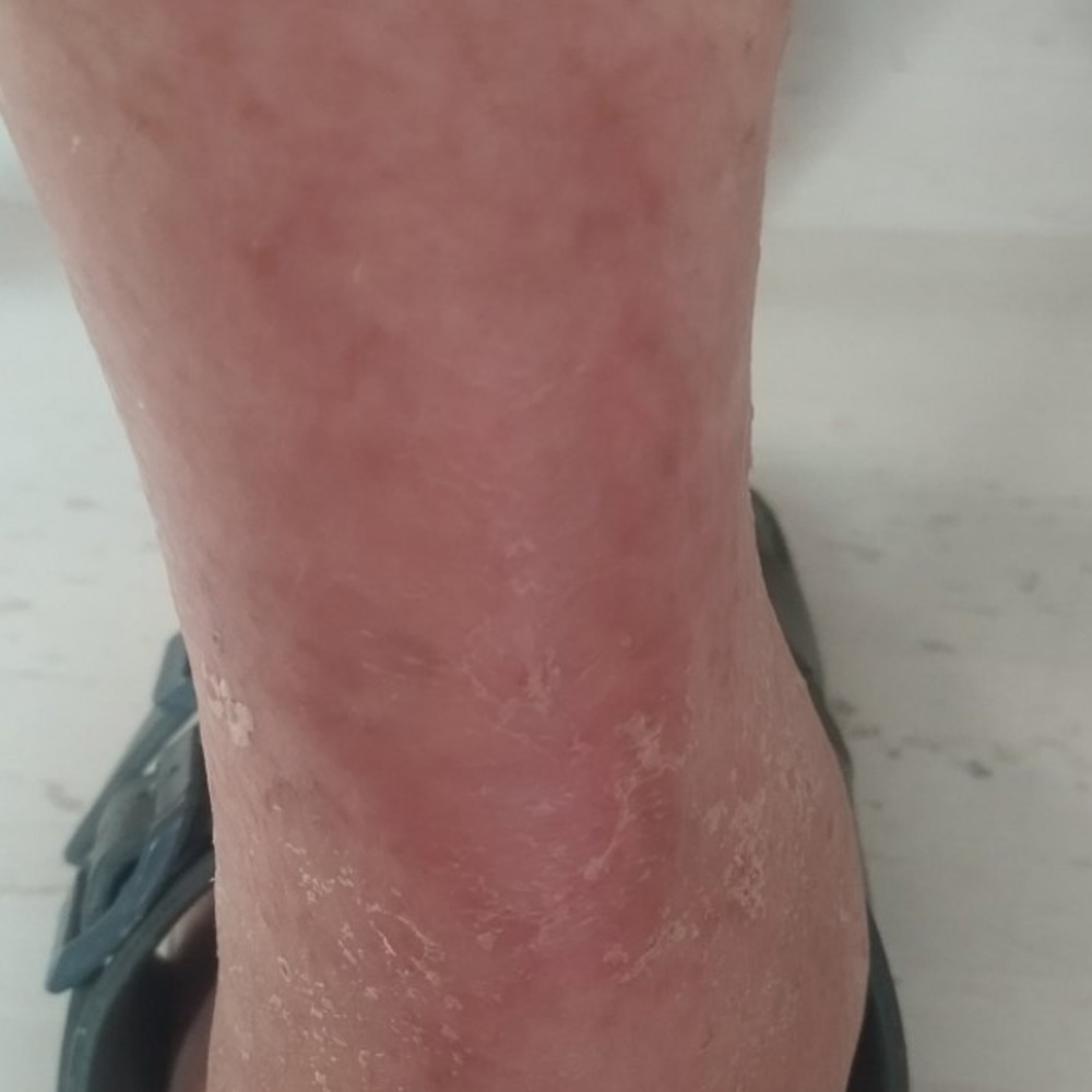
The wound achieved complete healing by secondary intention within approximately two months. This case illustrates that even ulcers in anatomically demanding locations can heal reliably when meticulous wound-bed preparation is combined with skin grafting as part of an advanced wound-healing strategy, restoring durable soft-tissue coverage and preserving limb function.
Related Clinical Cases
Concerned About a Diabetic Foot Problem?
Request a diabetic foot assessment and provide our team with the information needed to review your case.
Request a Diabetic Foot Assessment