
Diabetic Foot Tests: Essential Examinations for Diagnosis and Assessment
Diabetic Foot Assessment Before a Wound Develops Diabetic foot care... Read more
DIABETIC FOOT SURGERY · CLINICAL CASES
Wound-healing strategy depends on perfusion, infection control, tissue depth, pressure distribution and the condition of the postoperative wound bed.
Complex diabetic foot wounds often require coordinated infection control, careful wound-bed preparation and advanced methods that support granulation, tissue coverage and closure. These cases show how wound management complements diabetic foot treatment and the postoperative phase of a limb-salvage strategy.
For the complete collection organized by surgical approach, visit the Diabetic Foot Surgery – Clinical Cases hub.
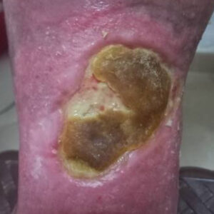
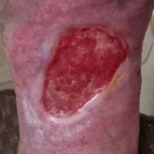
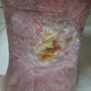
A 62-year-old woman with obesity presented with a chronic venous leg ulcer that had persisted for approximately four years despite prolonged conservative treatment. The ulcer remained painful, with extensive devitalized tissue and persistent inflammation, significantly limiting mobility and quality of life. The chronic nature of the wound made spontaneous healing highly unlikely without surgical intervention.
The ulcer was initially managed with meticulous surgical debridement to remove all non-viable tissue and create a healthy wound bed suitable for reconstruction. Once adequate granulation tissue had developed, a split-thickness skin graft was applied to achieve wound coverage. Throughout treatment, compression therapy with elastic bandaging was used to improve venous return, reduce edema, support graft integration, and promote optimal wound healing.
The wound progressively healed over approximately eight months, resulting in complete and durable closure. This case demonstrates that even long-standing venous ulcers can heal successfully when meticulous debridement, appropriate skin grafting, and sustained compression therapy are combined as part of a comprehensive limb-preservation strategy. The patient continues compression treatment to reduce the risk of recurrence and maintain long-term skin integrity.
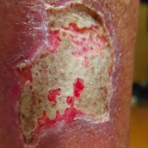
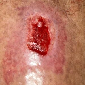
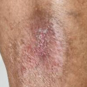
A 63-year-old man with type 2 diabetes presented with a superficial traumatic wound of the lower leg. Although the injury was not deep, healing was significantly delayed because of diabetes and the presence of extensive slough and bacterial biofilm covering the wound surface. The wound failed to progress through the normal phases of healing and required advanced wound management.
Initial treatment focused on conservative wound-bed preparation using specialized dressings to gradually remove slough and disrupt the biofilm while preserving healthy tissue. After achieving a clean, well-vascularized granulating wound bed, a split-thickness skin graft was applied to accelerate wound closure and restore skin coverage.
The wound healed completely by secondary intention over approximately two months, with successful graft integration and restoration of skin integrity. This case highlights the importance of meticulous wound-bed preparation before skin grafting, demonstrating how advanced wound-healing techniques can achieve reliable healing even in patients with diabetes and delayed wound repair.
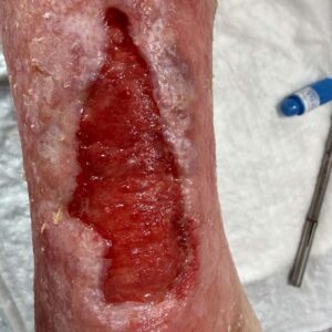
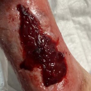
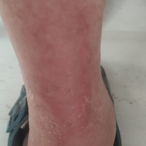
An 83-year-old woman with type 2 diabetes presented with a chronic ulcer located at the distal lower leg, extending toward the ankle—a region where wound healing is particularly challenging because of limited soft-tissue coverage, impaired vascularity, and constant mechanical stress. Despite local wound care, the ulcer failed to heal and required advanced surgical management.
Following meticulous surgical debridement, all devitalized tissue was removed to establish a healthy, well-vascularized wound bed. A split-thickness skin graft was then applied to promote definitive wound coverage and accelerate healing. Standard postoperative wound care and close clinical follow-up supported successful graft incorporation and progressive tissue regeneration.
The wound achieved complete healing by secondary intention within approximately two months. This case illustrates that even ulcers in anatomically demanding locations can heal reliably when meticulous wound-bed preparation is combined with skin grafting as part of an advanced wound-healing strategy, restoring durable soft-tissue coverage and preserving limb function.
Each case is presented to help advanced knowledge and improve patient outcomes.
NEED A CLINICAL EVALUATION?
Every diabetic foot problem requires individual assessment of infection, circulation, tissue viability and functional risk. Clinical cases are educational examples and do not replace an examination.
Diabetic Foot Assessment Before a Wound Develops Diabetic foot care... Read more

What Does “Diabetic Foot” Mean? Many people with diabetes have... Read more

Overview Limb-sparing surgery in the diabetic foot represents a modern... Read more