
Diabetic Foot Tests: Essential Examinations for Diagnosis and Assessment
Diabetic Foot Assessment Before a Wound Develops Diabetic foot care... Read more
DIABETIC FOOT SURGERY · CLINICAL CASES
Explore real osteomyelitis surgery cases showing how targeted bone surgery eradicates infection, preserves the foot, and often avoids major amputation.
Osteomyelitis surgery is guided by the extent of bone infection, soft-tissue involvement, perfusion and the possibility of preserving viable bone while achieving effective infection control.
Diabetic foot osteomyelitis requires careful assessment of infected bone and surrounding soft tissue. The cases below show how surgery can range from focused bone-preserving procedures to limited resection within a broader diabetic foot treatment plan and limb-salvage strategy.
For the complete collection organized by surgical approach, visit the Diabetic Foot Surgery – Clinical Cases hub.
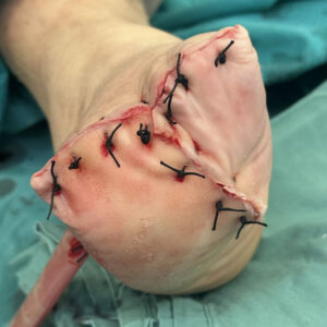
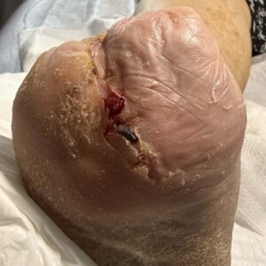
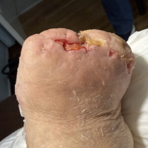
A 67-year-old patient with type 2 diabetes presented with osteomyelitis involving the second, third, and fourth metatarsals. Rather than performing multiple segmental metatarsal resections, a more functional limb-salvage strategy was selected to preserve foot stability and provide a durable weight-bearing surface.
A Lisfranc amputation was performed, removing the metatarsals and phalanges at the tarsometatarsal joint while preserving the tarsal bones. This approach created a balanced residual foot, offering better alignment and more stable contact with the ground than partial metatarsal resections.
The wound healed successfully following primary closure, with only minor superficial wound-healing issues that resolved during follow-up. The patient was subsequently fitted with a custom prosthetic filler and specialized therapeutic footwear, restoring a stable, plantigrade foot and enabling safe ambulation. This case demonstrates that, in selected patients with extensive metatarsal osteomyelitis, a Lisfranc amputation may provide superior long-term function compared with multiple isolated metatarsal resections.
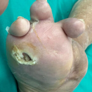
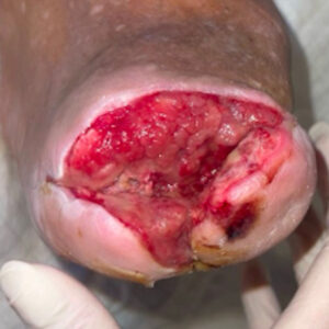
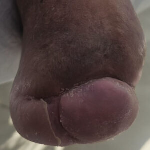
A 76-year-old man with type 2 diabetes was referred to our unit after previous amputation of the first and second rays at another hospital. He presented with a chronic plantar ulcer complicated by osteomyelitis of the fourth and fifth metatarsals, with extensive soft-tissue infection threatening further proximal spread and increasing the risk of major amputation.
The patient underwent a Lisfranc amputation, removing all infected bone and non-viable soft tissue until healthy margins were achieved. Because of the severity of the infection, the wound was intentionally left open rather than closed primarily, allowing adequate drainage and complete local infection control. Healing was planned by secondary intention, prioritizing eradication of the infection over immediate wound closure.
The wound healed completely over approximately three months, without recurrent infection or the need for further proximal amputation.
This approach preserved a functional residual foot suitable for ambulation with customized therapeutic footwear, demonstrating that even advanced forefoot osteomyelitis can often be managed successfully with a well-planned limb-salvage strategy.
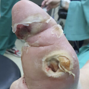
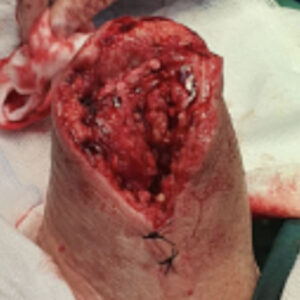
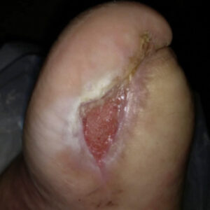
A 50-year-old man with type 2 diabetes was referred after previous amputation of the second, third, fourth, and fifth rays at another hospital. He subsequently developed osteomyelitis of the remaining first metatarsal, threatening complete loss of the forefoot and progression to a major lower-limb amputation.
Limb-salvage surgery was performed with complete excision of the infected first metatarsal and hallux until healthy, bleeding bone margins were achieved. The procedure was carefully planned to eradicate the infection while preserving a stable plantar stump capable of supporting weight-bearing with appropriate custom footwear.
The wound healed successfully, and the patient regained independent ambulation using a customized therapeutic shoe.
This case demonstrates that even when osteomyelitis affects the last remaining ray after previous multiple ray amputations, limb preservation may still be achievable. Rather than progressing to a major amputation, careful surgical planning can eradicate infection while creating a stable, functional residual foot that allows continued walking and preserves the patient’s independence.
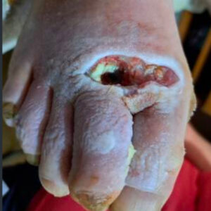
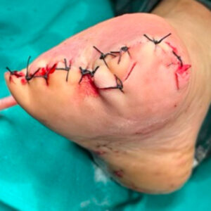
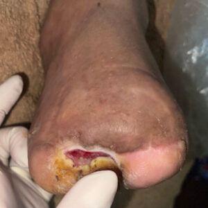
A 75-year-old man with type 2 diabetes was referred with an extensive diabetic foot infection complicated by a chronic ulcer and draining sinus tract. Imaging and intraoperative findings demonstrated osteomyelitis involving the first, second, and third metatarsals, making local bone preservation no longer feasible and placing the limb at high risk for major amputation.
Following complete assessment, a Lisfranc amputation was performed to eradicate all infected bone while preserving the maximum possible length of the foot. All infected tissues were excised until healthy, bleeding margins were obtained, and the plantar soft tissues were carefully preserved to create a durable, weight-bearing stump suitable for future ambulation.
The wound healed uneventfully, allowing preservation of a functional foot and avoiding a major lower-limb amputation.
This case demonstrates that, when forefoot osteomyelitis is too extensive for isolated bone resections, a well-planned minor amputation such as a Lisfranc amputation can provide definitive infection control while maintaining limb function. Preserving a stable, weight-bearing residual foot offers substantially better rehabilitation potential than a major amputation and should be considered whenever oncologically—or in this case infectiously—safe margins can be achieved.
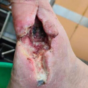
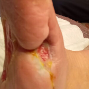
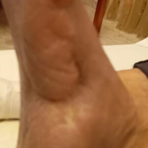
A 74-year-old man with type 2 diabetes was referred from another hospital after previous amputation of the first and second rays for diabetic foot osteomyelitis. Despite the earlier surgery, he developed recurrent osteomyelitis involving a tarsal bone, with persistent infection threatening further loss of the foot.
Limb-sparing surgery was performed with meticulous excision of the infected tarsal bone until healthy, bleeding bone margins were obtained. Every effort was made to preserve the remaining viable foot architecture and surrounding soft tissues, allowing eradication of the infection while maintaining the maximum possible structural stability and function.
The wound healed completely in approximately 3 months.
This case illustrates that recurrent diabetic foot osteomyelitis after previous partial foot amputations does not necessarily mandate a more proximal amputation. Careful identification and complete removal of the newly infected bone, while preserving all uninvolved structures, can successfully eradicate infection, preserve the remaining foot, and maintain functional ambulation.
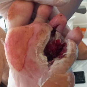
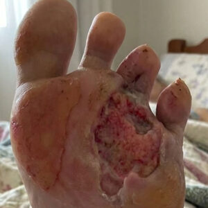
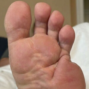
A 60-year-old patient with type 2 diabetes had previously undergone angioplasty for peripheral arterial disease, followed by amputation of the fifth toe and the proximal fifth metatarsal because of gangrene. Two years later, the patient returned with a plantar ulcer beneath the fourth metatarsal after failing to use the prescribed off-loading therapeutic footwear. Evaluation demonstrated osteomyelitis of the third and fourth metatarsals.
Surgical debridement was performed with resection of the distal portions of the third and fourth metatarsals, removing all infected bone while preserving as much of the foot as possible to maintain function.
The wound healed completely by secondary intention within three months. Following healing, the patient was fitted with custom off-loading footwear to reduce plantar pressure and prevent recurrent ulceration. This case highlights the critical importance of long-term pressure off-loading after minor amputations, as inadequate footwear can lead to recurrent ulcers, osteomyelitis, and the need for further bone resection.
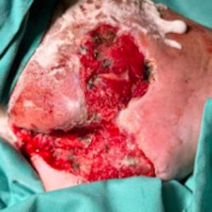
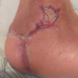
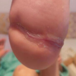
A 45-year-old woman with type 2 diabetes presented with calcaneal osteomyelitis after being advised at another hospital that major amputation was the only treatment option. The infection involved the calcaneus, threatening both limb preservation and independent ambulation.
Limb-sparing surgery was performed with meticulous excision of all infected calcaneal bone until healthy, bleeding bone margins were achieved. Every effort was made to preserve the surrounding viable heel soft tissues, maintaining the specialized plantar heel pad that is essential for durable weight-bearing and long-term foot function.
The wound healed completely in approximately 3.5 months, without the need for major amputation.
This case demonstrates that even calcaneal osteomyelitis—traditionally considered one of the most challenging diabetic foot infections—can often be successfully managed with aggressive bone-sparing surgery and preservation of the heel soft tissues. Careful eradication of infection, while maintaining the native weight-bearing heel pad, allowed limb salvage and preserved independent walking despite a previous recommendation for amputation.
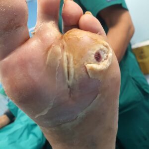
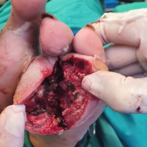
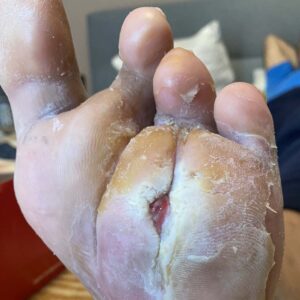
A 50-year-old man with type 2 diabetes, living abroad, presented with a chronic plantar ulcer of seven years’ duration. Despite prolonged conservative treatment, the wound failed to heal. Magnetic resonance imaging demonstrated osteomyelitis of the fourth metatarsal, identifying persistent bone infection as the underlying cause of the non-healing ulcer.
Limb-sparing surgery was performed with complete resection of the infected fourth metatarsal and the proximal phalanx of the fourth toe until healthy, bleeding bone margins were obtained. The surrounding viable plantar soft tissues were preserved to provide durable biological coverage of the surgical defect and facilitate healing by secondary intention.
Complete wound healing was achieved in approximately three months.
This case demonstrates that even a longstanding plantar ulcer of seven years can heal rapidly once the underlying osteomyelitis is surgically eradicated. Targeted bone resection eliminated the source of chronic infection, preserving a functional, weight-bearing foot while avoiding major amputation.
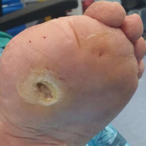
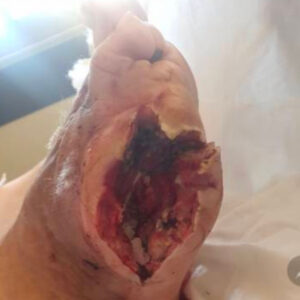
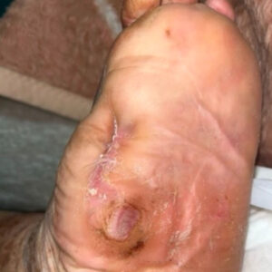
An elderly patient with type 2 diabetes was referred from another hospital following partial resection of the first metatarsal for diabetic foot osteomyelitis. Despite the initial surgery, persistent infection remained within the residual central portion of the first metatarsal, resulting in a chronic plantar wound with ongoing osteomyelitis.
Revision limb-sparing surgery was performed with complete excision of the residual infected first metatarsal until healthy, bleeding bone margins were achieved. Following meticulous debridement, negative pressure wound therapy (NPWT) was applied to promote granulation tissue formation, reduce wound volume, and prepare the wound for definitive healing.
Complete wound healing was achieved in approximately four months. This case illustrates that residual osteomyelitis after incomplete bone resection does not necessarily mandate major amputation. Careful revision surgery combined with NPWT can successfully eradicate persistent infection while preserving a functional, weight-bearing foot.
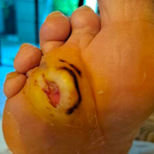
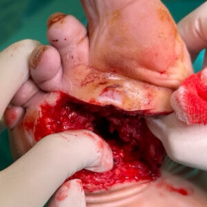
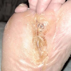
A 60-year-old woman with type 2 diabetes and peripheral arterial disease had previously undergone successful limb-sparing surgery by our team following lower-limb angioplasty for osteomyelitis of the second metatarsal. Two years later, she presented with a new plantar ulcer that had progressed to osteomyelitis involving the third metatarsal. The previous surgical scar, visible adjacent to the new lesion, demonstrates that this represented a new infectious episode rather than recurrence of the original infection.
Radical surgical debridement was performed with complete resection of the infected third metatarsal and the involved phalanges until healthy, bleeding bone margins were obtained. Viable plantar soft tissues were carefully preserved and mobilized to provide durable biological coverage of the resection cavity, maintaining forefoot stability and maximizing the potential for functional healing.
Complete wound healing was achieved in approximately three months.
Despite developing a second episode of metatarsal osteomyelitis two years after the initial limb-sparing procedure, the patient again avoided major amputation. Sequential conservative bone resections successfully eradicated infection while preserving a functional, weight-bearing foot, demonstrating that recurrent diabetic foot osteomyelitis can often be managed with repeat limb-salvage surgery rather than more proximal amputation.
Each case is presented to help advanced knowledge and improve patient outcomes.
NEED A CLINICAL EVALUATION?
Every diabetic foot problem requires individual assessment of infection, circulation, tissue viability and functional risk. Clinical cases are educational examples and do not replace an examination.
Diabetic Foot Assessment Before a Wound Develops Diabetic foot care... Read more

What Does “Diabetic Foot” Mean? Many people with diabetes have... Read more

Overview Limb-sparing surgery in the diabetic foot represents a modern... Read more