Residual First Metatarsal Osteomyelitis After Previous Surgery
Clinical presentation, surgical treatment and documented healing.
- 01 / Problem
- Clinical presentation
- 02 / Procedure
- Surgical approach
- 03 / Outcome
- Healing & follow-up
Clinical photographs below include wounds and surgical treatment.
01 / CLINICAL PRESENTATION
Problem
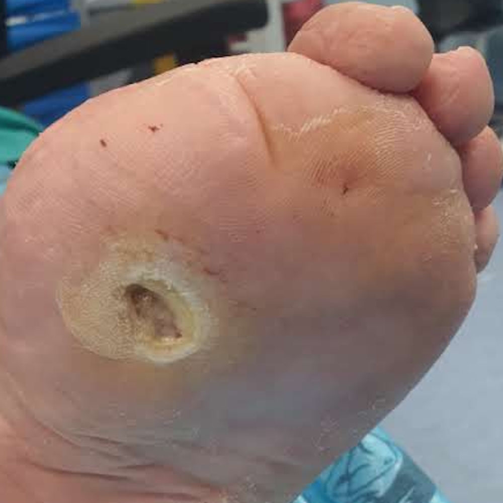
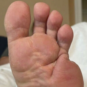
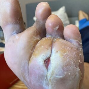
An elderly patient with type 2 diabetes was referred from another hospital following partial resection of the first metatarsal for diabetic foot osteomyelitis. Despite the initial surgery, persistent infection remained within the residual central portion of the first metatarsal, resulting in a chronic plantar wound with ongoing osteomyelitis.
02 / SURGICAL APPROACH
Procedure
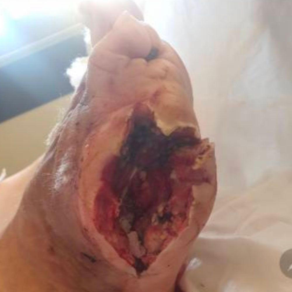
Revision limb-sparing surgery was performed with complete excision of the residual infected first metatarsal until healthy, bleeding bone margins were achieved. Following meticulous debridement, negative pressure wound therapy (NPWT) was applied to promote granulation tissue formation, reduce wound volume, and prepare the wound for definitive healing.
03 / HEALING & FOLLOW-UP
Outcome
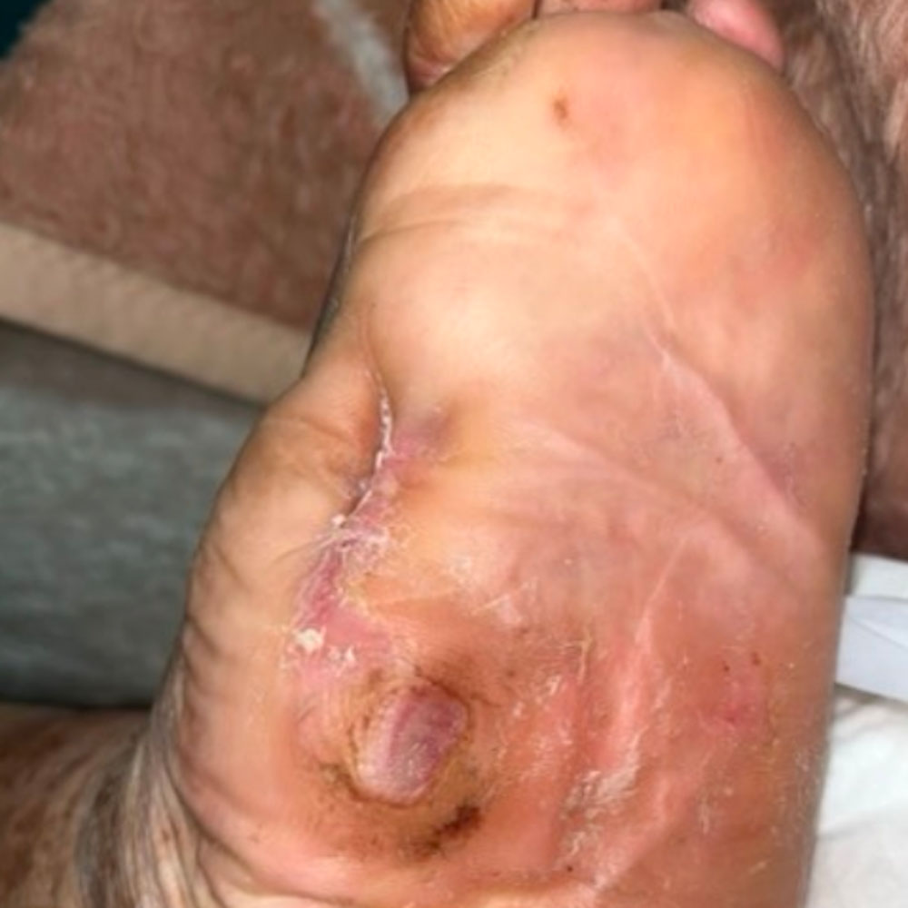
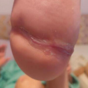
Complete wound healing was achieved in approximately four months. This case illustrates that residual osteomyelitis after incomplete bone resection does not necessarily mandate major amputation. Careful revision surgery combined with NPWT can successfully eradicate persistent infection while preserving a functional, weight-bearing foot.
Concerned About a Diabetic Foot Problem?
Request a diabetic foot assessment and provide our team with the information needed to review your case.
Request a Diabetic Foot Assessment