Diabetic Foot Limb Salvage Surgery: Advanced Surgical Treatment Options for Diabetic Foot Management
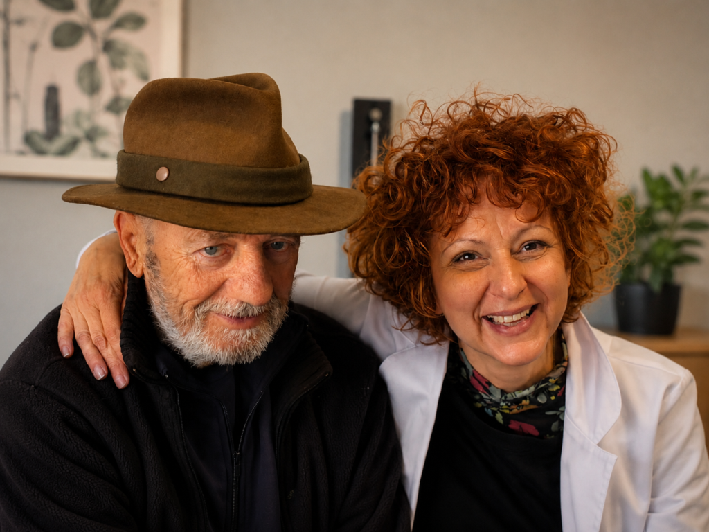
Treatment is tailored to the extent of diabetic foot tissue involvement, infection, vascular status, and structural damage, aiming to preserve foot function and support limb salvage.
Preserve Foot Function
Control Infection
Restore Blood Flow
Support Limb Salvage
Limb-Sparing Procedures
Treating diabetic foot while preserving the limb whenever safely possible

Preserve the Foot Whenever Possible

Control Infection
Restore Blood Flow

Support Limb Salvage
Frequently Asked Questions
1. What does limb-sparing surgery mean?
Limb-sparing surgery aims to treat infection and tissue damage while preserving as much of the foot as possible. The goal is to avoid major amputation whenever it is safe, maintaining a stable and functional foot that can support standing and walking.
2. Is limb-sparing always possible?
In many cases, yes. The decision depends on the extent of infection, the amount of viable tissue, and, most importantly, the level of blood flow (perfusion). When these conditions allow it, limb-sparing is always the preferred approach, as it offers better functional outcomes.
3. Will I still be able to walk after limb-sparing surgery?
Yes. Preserving the ability to stand and walk is a central goal of limb-sparing surgery. In most cases, patients are able to walk, often with appropriate support such as specialized footwear, insoles, or orthotics, depending on the extent of the procedure.
4. What role does blood flow play in limb preservation?
Adequate blood flow is essential for healing. Without sufficient perfusion, wounds cannot heal properly and surgical treatment may fail. In many patients, restoring circulation through revascularisation is a key step that makes limb-sparing surgery possible.
5. How is the treatment plan decided?
Treatment is individualized for each patient. It is based on a careful assessment of infection, tissue viability, and blood flow. The goal is to choose the safest approach that preserves as much function as possible while ensuring proper healing.
6. Are limb-sparing procedures painful?
No. These procedures are performed under local or regional anesthesia, often using nerve blocks, so patients do not feel pain during surgery. General anesthesia is usually not required.
7. Is hospitalization required after a limb-sparing procedure?
In many cases, hospitalization is short. Patients may return home the same day or the following day after surgery, depending on the extent of the procedure and their overall condition.
Types of Debridement & Drainage Surgery for Diabetic Ulcer
Removal of infected and non-viable tissue, with drainage of abscesses, to control infection and promote healing.
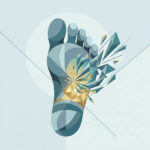
Surgical Debridement
Removal of non-viable and infected tissue. Sharp excision of devitalized tissue to expose healthy, viable tissue and restore healing potential.
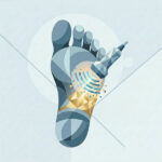
Ultrasound-Assisted Debridement
Selective removal with tissue preservation. Ultrasound-based debridement allows precise removal of non-viable tissue while preserving viable, well-perfused structures.
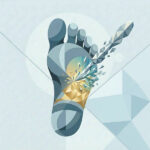
Incision & Drainage
Control of deep infection and abscess. Opening of infected spaces to release pus and reduce pressure, preventing further spread of infection.
Frequently Asked Questions
1. What is a diabetic foot ulcer?
A diabetic foot ulcer is an open sore or wound on the foot of a person with diabetes, most often on weight-bearing areas such as the sole or toes.
It develops due to nerve damage (loss of protective sensation) and/or reduced blood flow (poor circulation), making the skin more vulnerable to injury and delaying healing.
2. Why do these ulcers become infected?
Because they are open wounds, bacteria can easily enter the tissue. In diabetes, infection is more likely due to loss of sensation (neuropathy), reduced blood flow (poor perfusion), and impaired immune response. Deep or long-standing ulcers carry an even higher risk. Without timely treatment, many of these wounds can progress to infection.
3. What is debridement in a diabetic foot ulcer?
Debridement is a procedure in which dead, infected, or damaged tissue, as well as surrounding callus, is removed, leaving healthy, well-perfused tissue behind.
It helps control infection, improve the wound environment, and allows accurate assessment of the ulcer’s depth and extent.
4. What is drainage in a diabetic foot ulcer?
Drainage is a procedure in which the surgeon opens and releases pus or infected fluid from deep spaces around the ulcer. In some cases, a small drain is placed to allow continued evacuation and proper cleaning.
It is used when infection spreads beneath the skin, forming abscesses or fluid collections, and is usually combined with debridement, antibiotics, and off-loading as part of limb-preserving treatment.
5. Can antibiotics alone treat a diabetic foot infection?
Sometimes, in mild and superficial infections without abscess or bone involvement.
In deeper or more severe infections—especially with abscess, necrotic tissue, or osteomyelitis—antibiotics alone are not enough. Surgical debridement and/or drainage are usually required, often combined with off-loading and optimization of perfusion.
6. What role does perfusion play in healing?
Perfusion refers to the blood supply reaching the tissues. Adequate perfusion is essential for healing, as blood delivers oxygen, nutrients, and immune cells needed for tissue repair and infection control.
When perfusion is poor, wounds heal slowly or not at all, are more prone to infection, and carry a higher risk of amputation. Assessing tissue perfusion helps guide treatment decisions, including the need for revascularization.
7. What should patients expect after debridement or drainage?
Recovery focuses on wound healing and infection control. Regular follow-up, dressing changes, and off-loading are essential to support healing.
In some cases, repeated debridement may be needed. Ongoing monitoring helps detect early signs of recurrence and guides further treatment.
8. Is hospitalization or general anesthesia required for debridement or drainage?
In most cases, no. These procedures are typically performed under local or regional anesthesia, including nerve blocks, so there is no pain during the procedure. Hospitalization is usually short, and many patients can return home the same day or the following day.
Types of Osteomyelitis Surgery
Targeted resection of infected bone with preservation of viable, well-perfused tissue.

Phalangeal Surgery (Toe Phalanx Resection)
Preserving the toe whenever possible Removal of infected phalanges while maintaining the soft-tissue envelope, preserving toe length and function when perfusion allows.

Metatarsal Surgery (Partial or Complete Resection)
Maintaining foot stability while removing infection Partial or complete resection of the affected metatarsal to control infection while preserving surrounding structures and foot mechanics.

Tarsal Bone Resection
Targeted treatment with maximal tissue preservation Focused removal of infected tarsal bone, preserving adjacent joints and structures when viable and well perfused.

Calcaneal Surgery (Partial Calcanectomy)
Partial resection of the calcaneus to control infection and preserve a functional, weight-bearing limb whenever adequate soft-tissue coverage and perfusion are present.

Transmetatarsal Procedure
Preserving a stable, weight-bearing foot Removal of the forefoot at the metatarsal level to control infection while maintaining a functional, weight-bearing limb.

Lisfranc and Chopart Procedures
Managing advanced infection while preserving function Limb-sparing partial foot procedures used in severe cases to remove infection while maintaining the ability to stand and mobilize.
Frequently Asked Questions
1. Can a simple foot wound reach the bone?
Yes. In diabetes, infections can spread from a wound to the bone, leading to osteomyelitis. Early evaluation is essential and can help save the foot.
2. How do we know if a foot wound has reached the bone?
It is determined through clinical examination and, when needed, imaging such as X-ray or MRI. These help assess whether the infection has reached the bone, a condition known as osteomyelitis.
3. What tests are needed for a diabetic foot?
Evaluation usually starts with an X-ray to assess possible bone involvement. In earlier stages, Magnetic Resonance Imaging (MRI) may be needed to detect osteomyelitis and map the extent of infection, helping guide the extent of bone resection. Early and appropriate investigation supports timely treatment.
4. Can the foot be saved in a patient with diabetes?
In many cases, yes. Even in the presence of severe infection or osteomyelitis, surgery can control the infection while preserving foot function. Early intervention is key to avoiding amputation.
5. What happens before surgery for osteomyelitis?
Before surgery, careful planning is essential. Evaluation usually includes X-ray and, when needed, Magnetic Resonance Imaging (MRI) to define the extent of infection. These findings guide the surgical plan, including which bone and soft tissue need to be removed. The patient is fully informed before the procedure.
6. What is done during surgery for osteomyelitis in the diabetic foot?
The goal is to remove the infection while preserving foot function. During surgery, infected and non-viable tissue, including the affected bone, is carefully removed while preserving well-perfused, healthy tissue. Tissue samples are taken to guide antibiotic therapy. The aim is a stable, functional foot.
7. Does osteomyelitis always mean amputation?
No. In many cases, targeted bone resection can control the infection while preserving the limb. Adequate perfusion is essential for healing and guides surgical decision-making.
8. Is surgery for osteomyelitis in the diabetic foot painful?
No. The procedure is performed under local or regional anesthesia, including nerve blocks, so there is no pain during surgery. General anesthesia is usually not required.
9. Is hospitalization required?
In many cases, hospitalization is short. Patients may return home the same day or the following day after surgery. The procedure is usually simpler than expected.
10. How do we choose the right antibiotics after surgery?
During surgery, samples are taken from bone and surrounding tissues to identify the causative organism. This allows for targeted antibiotic therapy, tailored to the severity and extent of the infection.
11. What happens after surgery for the diabetic foot?
Postoperative care focuses on wound healing and infection control. Regular follow-up and, when needed, wound debridement are essential to monitor progress and detect early signs of recurrence. Remote assessment through wound images and, when required, home visits by a specialized nursing team support continuous, individualized care.
12. What should be considered after surgery for the diabetic foot?
Protecting the foot is essential for healing. Off-loading, using specialized footwear or immobilization devices, reduces pressure on the wound. Good glycemic control and overall health also play a key role in safe and effective recovery.
13. How is osteomyelitis in the diabetic foot treated overall?
Osteomyelitis requires early diagnosis and a coordinated approach. Treatment combines targeted antibiotics, surgery when needed, proper off-loading, and close follow-up. A multidisciplinary team is essential to achieve optimal outcomes and preserve the limb.
Without proper coordination, the risk of complications and limb loss increases.
Minor Amputations
Includes toe, ray, transmetatarsal, Lisfranc, and Chopart amputations based on infection control and adequate perfusion, aiming to preserve foot length, stability and function.

Toe amputation
Removal of one toe, or part of a toe, when infection or tissue damage is confined to that area. This procedure preserves as much foot structure as possible and usually has the least impact on walking and balance.

Ray amputation
Removal of a toe together with part or all of its corresponding metatarsal bone when infection or osteomyelitis extends beyond the toe. This procedure aims to eradicate infection while preserving foot stability, function, and weight-bearing ability whenever possible.

Partial Calcanectomy
Preserving a functional, weight-bearing limb: partial resection of the calcaneus to remove non-viable bone and tissue while preserving limb stability and the ability to bear weight whenever adequate perfusion and soft-tissue coverage are present.

Transmetatarsal amputation (TMA)
Removal of the forefoot across the metatarsal bones when infection, osteomyelitis, or non-viable tissue involves multiple toes or a larger area. The procedure preserves the hindfoot, aiming to maintain standing, walking, and a stable, functional weight-bearing limb whenever possible.

Lisfranc amputation and Chopart amputation
Partial foot amputations performed when infection, osteomyelitis, or extensive tissue loss extends beyond the forefoot. By preserving as much of the midfoot and hindfoot as possible, these procedures aim to maintain limb stability, weight-bearing capacity, and functional ambulation whenever feasible.
Frequently Asked Questions
1. What type of anesthesia is used for minor amputations?
Minor amputations are usually performed under local or regional anesthesia, including nerve blocks, so there is no pain during the procedure. General anesthesia is typically not required.
2. How long will I stay in the hospital after a minor amputation?
Hospital stay is usually short, often around 24 hours. In many cases, patients are discharged the next day, depending on their overall condition and recovery.
3. How much of the foot will be removed?
Only the part that cannot be saved is removed. The goal is to preserve as much length and function as possible while fully controlling the infection.
4. Will I be able to walk after a minor amputation?
Most patients can walk again. The level of amputation determines whether simple footwear or custom support is needed.
5. When will I be able to walk after a minor amputation?
In most cases, walking starts early, often from the first day after surgery using a specialized postoperative shoe. Further progression depends on wound healing and the level of amputation.
6. Will I need special footwear after surgery?
Yes. Most patients require custom footwear, insoles, or prosthetic support to protect the foot and improve walking, depending on the level of amputation.
Advanced Wound Healing Options
Uses advanced therapies such as grafts, ultrasound-assisted debridement and PRP to support healing and prepare the wound for closure
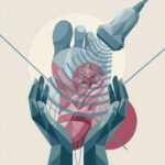
Ultrasound‑Assisted Wound Debridement (UAWD)
UAWD is a form of ultrasound‑assisted wound debridement used to clean the ulcer in an atraumatic, tissue‑sparing way. It uses low‑frequency ultrasound energy in a fluid medium to break up and remove biofilm, slough and non‑viable tissue, without cutting and without damaging healthy granulation tissue.

Collagen–Elastin Matrix Graft
Collagen–Elastin Matrix Graft A soft, biocompatible scaffold placed into the wound that acts as a framework for new tissue. It supports cell growth, blood vessel formation, and stable skin regeneration in deep or complex ulcers.

Platelet-Rich Plasma (PRP)
A small amount of the patient’s own blood is processed to concentrate growth factors and applied to the wound. It helps stimulate healing, improve tissue regeneration, and support wound closure.
Frequently Asked Questions
1. Where are these treatments performed?
These treatments can be performed either in an outpatient hospital setting or in our clinic, depending on the patient’s condition and the type of procedure. Most of them do not require hospital admission and are part of a structured outpatient treatment plan.
2. Do these treatments require anesthesia?
Most advanced wound healing treatments do not require anesthesia. In selected cases, such as ultrasound-assisted debridement, local anesthesia may be used to ensure comfort during the procedure.
3. Are these treatments painful?
These procedures are generally well tolerated and minimally invasive. Most patients experience little to no discomfort during treatment, especially when appropriate preparation and care are provided.
4. How many sessions are needed?
The number of sessions varies depending on the size, depth, and overall condition of the wound. Treatment is individualized and adjusted based on how the wound responds over time.
5. Do these treatments replace surgery?
No. These therapies are used as part of a comprehensive treatment strategy, alongside debridement, infection control, and proper wound care, to enhance healing and prepare the wound for closure.
6. How do these treatments support healing?
They improve the local wound environment, promote tissue regeneration, and support the formation of healthy, well-vascularized tissue, making wound closure more effective and stable.
Major Amputations (when necessary)
Performed only when the limb cannot be safely preserved, prioritizing patient safety and quality of life.

In some cases, when infection or tissue damage is too advanced, amputation above or below the knee may be necessary. We approach this decision with great care and empathy, always aiming to ensure the safest outcome and the best possible functional recovery for the patient.
In these cases, surgery is performed under general anesthesia, often combined with regional anesthesia. The regional block can provide pain control for the first few days after surgery, helping ensure a more comfortable recovery.
Frequently Asked Questions
1. Will I be in pain after surgery?
We take postoperative pain very seriously. With the use of combined anesthesia techniques, including regional blocks, pain is usually well controlled during the first days after surgery. Our goal is to keep you as comfortable as possible throughout your recovery.
2. Why would a major amputation be necessary?
A major amputation is considered only when the limb cannot be safely preserved. This is always a difficult decision, and it is made with great care, prioritizing your safety, your overall health, and your long-term quality of life.
3. How long will I stay in the hospital?
Hospital stay is usually a few days (about three days), depending on your condition and recovery. During this time, we closely monitor your progress and support you through the early stages after surgery.
4. Will I be able to walk again?
Yes. With proper rehabilitation, physiotherapy, and the use of a prosthesis when needed, patients can regain mobility. We guide and support you through each step of this process.
5. How do you support patients after such a surgery?
Recovery does not end with the operation. We provide continuous follow-up, guidance, and coordination with rehabilitation and prosthetic teams. Our aim is to help you adapt, recover, and regain independence.