Tarsal Osteomyelitis Following Previous Partial Foot Amputation
Clinical presentation, surgical treatment and documented healing.
- 01 / Problem
- Clinical presentation
- 02 / Procedure
- Surgical approach
- 03 / Outcome
- Healing & follow-up
Clinical photographs below include wounds and surgical treatment.
01 / CLINICAL PRESENTATION
Problem
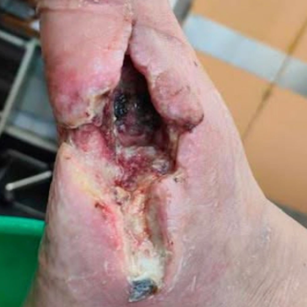
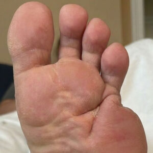
A 74-year-old man with type 2 diabetes was referred from another hospital after previous amputation of the first and second rays for diabetic foot osteomyelitis. Despite the earlier surgery, he developed recurrent osteomyelitis involving a tarsal bone, with persistent infection threatening further loss of the foot.
02 / SURGICAL APPROACH
Procedure
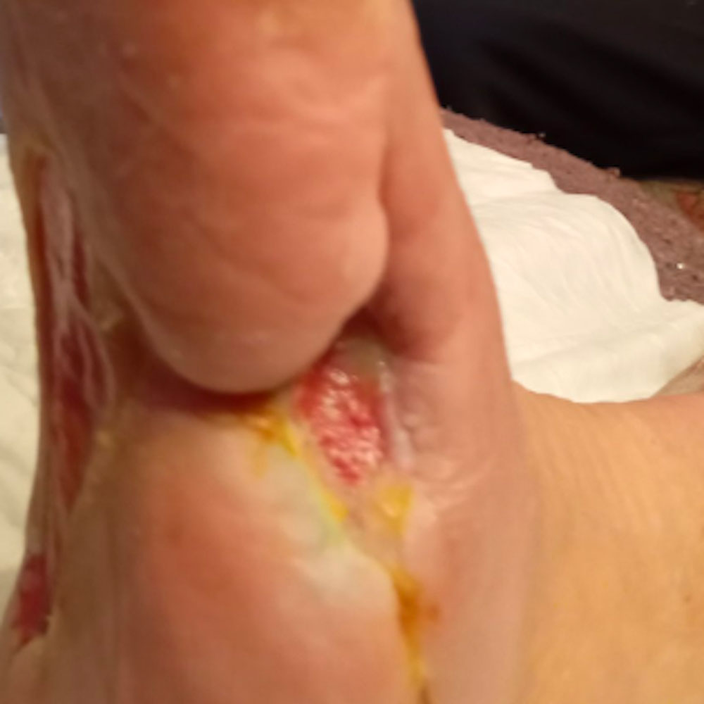
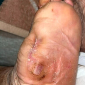
Limb-sparing surgery was performed with meticulous excision of the infected tarsal bone until healthy, bleeding bone margins were obtained. Every effort was made to preserve the remaining viable foot architecture and surrounding soft tissues, allowing eradication of the infection while maintaining the maximum possible structural stability and function.
03 / HEALING & FOLLOW-UP
Outcome
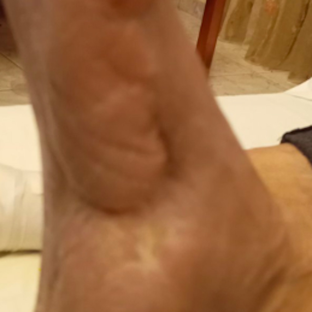
The wound healed completely in approximately 3 months.
This case illustrates that recurrent diabetic foot osteomyelitis after previous partial foot amputations does not necessarily mandate a more proximal amputation. Careful identification and complete removal of the newly infected bone, while preserving all uninvolved structures, can successfully eradicate infection, preserve the remaining foot, and maintain functional ambulation.
Concerned About a Diabetic Foot Problem?
Request a diabetic foot assessment and provide our team with the information needed to review your case.
Request a Diabetic Foot Assessment